Accessory spleen
| Accessory spleen | |
|---|---|
|
Histologic section of an accessory spleen | |
| Classification and external resources | |
| Specialty | medical genetics |
| ICD-10 | Q89.0 |
| ICD-9-CM | 759.0 |
| DiseasesDB | 32864 |
| eMedicine | article/896865 |
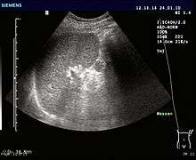
An accessory spleen (supernumerary spleen, splenule, or splenunculus) is a small nodule of splenic tissue found apart from the main body of the spleen. Accessory spleens are found in approximately 10 percent of the population[1] and are typically around 1 centimeter in diameter. They may resemble a lymph node or a small spleen. They form either by the result of developmental anomalies or trauma.[2] They are medically significant in that they may result in interpretation errors in diagnostic imaging[2] or continued symptoms after therapeutic splenectomy.[1]
Causes and locations
| Accessory spleen | |
|---|---|
| Details | |
| Identifiers | |
| Latin | splen accessorius, lien accessorius |
| TA | A13.2.01.022 |

Accessory spleens may be formed during embryonic development when some of the cells from the developing spleen are deposited along the path from the midline, where the spleen forms, over to its final location on the left side of the abdomen by the 9th–11th ribs. The most common locations for accessory spleens are the hilum of the spleen and adjacent to the tail of the pancreas. They may be found anywhere along the splenic vessels, in the gastrosplenic ligament, the splenorenal ligament, the walls of the stomach or intestines, the pancreatic tail,[3] the greater omentum, the mesentery or the gonads and their path of descent.[4] The typical size is approximately 1 centimeter, but sizes ranging from a few millimeters up to 2–3 centimeters are not uncommon.[2]
Splenogonadal fusion can result in one or more accessory spleens along a path from the abdomen into the pelvis or scrotum. The developing spleen forms near the urogenital ridge from which the gonads develop. The gonads may pick up some tissue from the spleen, and as they descend through the abdomen during development, they can produce either a continuous or a broken line of deposited splenic tissue.[4]
Splenosis is a condition where foci of splenic tissue undergo autotransplantation, most often following physical trauma or splenectomy. Displaced tissue fragments can implant on well vascularized surfaces in the abdominal cavity, or, if the diaphragmatic barrier is broken, the thorax.[5][6]
Significance
If splenectomy is performed for conditions in which blood cells are sequestered in the spleen, failure to remove accessory spleens may result in the failure of the condition to resolve.[1] During medical imaging, accessory spleens may be confused for enlarged lymph nodes or neoplastic growth in the tail of the pancreas,[3] gastrointestinal tract, adrenal glands or gonads.[2]
References
- 1 2 3 Moore, Keith L. (1992). Clinically Oriented Anatomy (3rd ed.). Baltimore: Williams & Wilkins. p. 187. ISBN 0-683-06133-X.
- 1 2 3 4 Gayer G; Zissin R; Apter S; Atar E; Portnoy O; Itzchak Y (August 2001). "CT findings in congenital anomalies of the spleen". British Journal of Radiology. British Institute of Radiology. 74 (884): 767–772. PMID 11511506. Retrieved 2009-03-03.
- 1 2 Kim SH; Lee JM; Han JK; Lee JY; Kim KW; Cho KC; Choi BI (March–April 2008). "Intrapancreatic Accessory Spleen: Findings on MR Imaging, CT, US and Scintigraphy, and the Pathologic Analysis". Korean J Radiol. Korean Radiological Society. 9 (2): 162–174. doi:10.3348/kjr.2008.9.2.162. PMC 2627219
 . PMID 18385564.
 - 1 2 Chen S–L; Kao Y–L; Sun H–S; Lin W–L (November 2008). "Splenogonadal Fusion". Journal of the Formosan Medical Association. 107 (11): 892–5. doi:10.1016/S0929-6646(08)60206-5. ISSN 0929-6646. PMID 18971159. Retrieved 2009-03-03.
- ↑ Abu Hilal M; Harb A; Zeidan B; Steadman B; Primrose JN; Pearce NW (January 5, 2009). "Hepatic splenosis mimicking HCC in a patient with hepatitis C liver cirrhosis and mildly raised alpha feto protein; the important role of explorative laparoscopy". World Journal of Surgical Oncology. England: BioMed Central. 7 (1): 1. doi:10.1186/1477-7819-7-1. PMC 2630926
 . PMID 19123935.
 - ↑ Madjar S; Weissberg D (October 1994). "Thoracic splenosis". Thorax. British Medical Assn. 49 (10): 1020–1022. doi:10.1136/thx.49.10.1020. ISSN 0040-6376. PMC 475241
 . PMID 7974296.