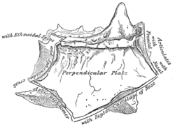
Perpendicular plate of ethmoid bone
| Perpendicular plate of ethmoid bone | |
|---|---|
 | |
 Ethmoid bone from behind. | |
| Details | |
| Identifiers | |
| Latin | lamina perpendicularis ossis ethmoidalis |
| TA | A02.1.07.006 |
| FMA | 52891 |

The perpendicular plate of the ethmoid bone (vertical plate) is a thin, flattened lamina, polygonal in form, which descends from the under surface of the cribriform plate, and assists in forming the septum of the nose; it is generally deflected a little to one or other side. The anterior border articulates with the spine of the frontal bone and the crest of the nasal bones.
The posterior border articulates by its upper half with the sphenoidal crest, by its lower with the vomer.
The inferior border is thicker than the posterior, and serves for the attachment of the septal nasal cartilage of the nose.
The surfaces of the plate are smooth, except above, where numerous grooves and canals are seen; these lead from the medial foramina on the cribriform plate and lodge filaments of the olfactory nerves.
Additional images
 Medial wall of left nasal fossa.
 Bones and cartilages of septum of nose. Right side.
- Perpendicular plate of Ethmoid.
References
This article incorporates text in the public domain from the 20th edition of Gray's Anatomy (1918)
External links
- Anatomy figure: 33:02-02 at Human Anatomy Online, SUNY Downstate Medical Center - "Diagram of skeleton of medial (septal) nasal wall."
- Atlas image: rsa1p7 at the University of Michigan Health System - "Nasal septum, lateral view"
- lesson9 at The Anatomy Lesson by Wesley Norman (Georgetown University) (nasalseptumbonescarti)