Vulvar intraepithelial neoplasia
| Vulvar intraepithelial neoplasia | |
|---|---|
 | |
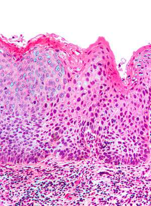
| Micrograph of (classic) vulvar intraepithelial neoplasia III. H&E stain. | |
| Classification and external resources | |
| Specialty | oncology |
| ICD-10 | D07.1 (ILDS D07.120) |
| ICD-9-CM | 233.32 |
The term Vulvar intraepithelial neoplasia (VIN) refers to particular changes that can occur in the skin that covers the vulva. VIN is not cancer, and in some women it disappears without treatment. If the changes become more severe, there is a chance that cancer might develop after many years, and so it is referred to as a precancerous condition.[1]
ISSVD Classification
Medically speaking, the term denotes a squamous intraepithelial lesion of the vulva that shows dysplasia with varying degrees of atypia. The epithelial basement membrane is intact and the lesion is thus not invasive but has invasive potential.
The terminology of VIN evolved over several decades. In 1989[2] the Committee on Terminology, International Society for the Study of Vulvar Disease (ISSVD) replaced older terminology such as vulvar dystrophy, Bowen's disease, and Kraurosis vulvae by a new classification system for Epithelial Vulvar Disease:
- Nonneoplastic epithelial disorders of vulva and mucosa:
- Lichen sclerosus
- Squamous hyperplasia
- Other dermatoses
- Mixed neoplastic and nonneoplastic disorders
- Intraepithelial neoplasia
- Squamous vulvar intraepithelial neoplasia (VIN)
- VIN I, mildest form
- VIN II, intermediate
- VIN III, most severe form including carcinoma in situ of the vulva
- Non-squamous intraepithelial neoplasia
- Extramammary Paget's disease
- Tumors of melanocytes, noninvasive
- Squamous vulvar intraepithelial neoplasia (VIN)
- Invasive disease (vulvar carcinoma)
The ISSVD further revised this classification in 2004, replacing the three-grade system with a single-grade system in which only the high-grade disease is classified as VIN. This is subdivided into: 1) usual-type VIN (including warty, basaloid and mixed subtypes)commonly associated with carcinogenic genotypes of HPV and/or HPV persistence factors such as cigarette smoking or immunocompromised states and 2) differentiated VIN, commonly associated with vulvar dermatoses such as lichen sclerosis. Differentiated VIN associated with lichen sclerosis, however, is more likely to be associated with squamous carcinoma than is usual-type VIN.[3] [4]
Causes
The exact cause of VIN is unknown. Studies are being done to determine the cause of VIN. The following factors have been associated with VIN:
- HPV (Human Papilloma Virus)
- HSV-2 (Herpes simplex Virus - Type 2)
- Smoking
- Immunosuppression
- Chronic vulvar irritation
- Conditions such as Lichen Sclerosus
Diagnosis


The patient may have no symptoms, or local symptomatology including itching, burning, and pain. The diagnosis is always based on a careful inspection and a targeted biopsy of a visible vulvar lesion.
The type and distribution of lesions varies among the two different types of VIN. In the Usual type VIN, seen more frequently in young patients, lesions tend to be multifocal over an otherwise normal vulvar skin. In the differentiated type VIN, usually seen in postmenopausal women, lesions tend to be isolated and are located over a skin with a vulvar dermatosis such as Lichen slerosus.[3]
Prevention
Vaccinating girls with HPV vaccine before their initial sexual contact has been claimed to reduce incidence of VIN.[5]
Additional images
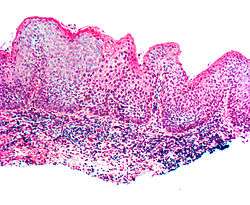
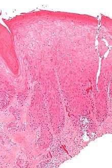
.jpg) Micrograph of vulvar intraepithelial neoplasia.
Micrograph of vulvar intraepithelial neoplasia.
References
- ↑ "Vulval intra-epithelial neoplasia (VIN)". Macmillan Cancer Support. Retrieved 2010-06-09.
- ↑ Ridley CM, Frankman O, Jones IS, et al. (May 1989). "New nomenclature for vulvar disease: International Society for the Study of Vulvar Disease". Hum. Pathol. 20 (5): 495–6. doi:10.1016/0046-8177(89)90019-1. PMID 2707802.
- 1 2 http://www.issvd.org
- ↑ Sideri M, Jones RW, Wilkinson EJ, Preti M, Heller D,. Scurry J, Haefner H, Neill S. 2004 Modified Terminology, ISSVD Vulvar Oncology Subcommittee. Journal of Reproductive Medicine. 2005;50:807-10.
- ↑ "FDA Approves Expanded Uses for Gardasil to Include Preventing Certain Vulvar and Vaginal Cancers". 2008-09-12. Retrieved 2010-02-13.