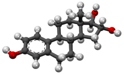
Estriol
 | |
 | |
| Clinical data | |
|---|---|
| Trade names | Ovestin[1] |
| Routes of administration | Oral, vaginal |
| ATC code |
G03CA04 (WHO) G03CC06 (WHO) |
| Pharmacokinetic data | |
| Biological half-life | 5 hours[2] |
| Identifiers | |
| Synonyms | 16α-Hydroxyestradiol; Estra-1,3,5(10)-triene-3,16α,17β-triol |
| CAS Number |
50-27-1 |
| PubChem (CID) | 5756 |
| IUPHAR/BPS | 2821 |
| DrugBank |
DB04573 |
| ChemSpider |
5553 |
| UNII |
FB33469R8E |
| KEGG |
C05141 |
| ChEBI |
CHEBI:27974 |
| ChEMBL |
CHEMBL193482 |
| Chemical and physical data | |
| Formula | C18H24O3 |
| Molar mass | 288.39 g·mol−1 |
| 3D model (Jmol) | Interactive image |
| |
| |
| (verify) | |
Estriol (E3), or oestriol, also known as 16α-hydroxyestradiol or as estra-1,3,5(10)-triene-3,16α,17β-triol, is a natural steroidal estrogen and one of the three main estrogens in the human body.[3] Production of estriol by the ovaries is almost undetectable in non-pregnant women.[4] However, during pregnancy, estriol is synthesized in very high amounts by the placenta and is by far the most produced estrogen in the body,[4][5] although circulating levels are similar to those of other estrogens due to a relatively high rate of metabolism and excretion.[5][6]
Biosynthesis
During pregnancy
Estriol is produced in notable quantities only during pregnancy.[4] Levels of estriol increase 1,000-fold during pregnancy,[7] whereas levels of estradiol and estrone increase 100-fold,[8] and estriol accounts for 90% of the estrogens in the urine of pregnant women.[6] At term, the daily production of estriol by the placenta is 35 to 45 mg,[8] and levels in the maternal circulation are 8 to 13 ng/dL.[4]
The placenta produces pregnenolone and progesterone from circulating cholesterol.[5] Pregnenolone is taken up by the fetal adrenal glands and converted into dehydroepiandrosterone (DHEA), which is then sulfated by steroid sulfotransferase into dehydroepiandrosterone sulfate (DHEA-S). DHEA-S is hydroxylated by high CYP3A7 expression and activity into 16α-hydroxy-DHEA-S (16α-OH-DHEA-S) in the fetal liver and to a limited extent in the fetal adrenal glands.[4][9] 16α-OH-DHEA-S is then taken up by the placenta.[4] Due to high expression of steroid sulfatase in the placenta, 16α-OH-DHEA-S is rapidly cleaved into 16α-OH-DHEA.[4] Then, 16α-OH-DHEA is converted by 3β-hydroxysteroid dehydrogenase type I (3β-HSD1) into 16α-hydroxyandrostenedione (16α-OH-A4) and 16α-OH-A4 is converted by aromatase into 16α-hydroxyestrone (16α-OH-E1),[10] which is subsequently converted into estriol by 17β-hydroxysteroid dehydrogenase and then secreted predominantly into the maternal circulation.[4][11] Approximately 90% of precursors in estriol formation originate from the fetus.[11]
During pregnancy, 90 to 95% of estriol in the maternal circulation is conjugated in the form of estriol glucuronide and estriol sulfate, and levels of unconjugated estriol are slightly less than those of unconjugated estradiol and similar to those of unconjugated estrone.[6] As such, target tissues are likely to be exposed to similar amounts of free estriol, estradiol, and estrone during pregnancy.[6]
Estrone and estradiol are also produced in the placenta during pregnancy.[4] However, in the case of estrone and estradiol, DHEA-S is taken up by the placenta and cleaved by steroid sulfatase into dehydroepiandrosterone (DHEA), DHEA is converted by 3β-hydroxysteroid dehydrogenase type I into androstenedione, and androstenedione is aromatized into estrone.[4] Then, placental 17β-hydroxysteroid dehydrogenase interconverts estrone and estradiol and the two hormones are secreted into the maternal circulation.[4] DHEA-S that is taken up by the placenta is mainly produced by the fetal adrenal glands.[4]
Estetrol, which is the 15α-hydroxylated derivative of estriol, is synthesized from 15α-OH-DHEA-S (which is produced in the fetal liver) during pregnancy.[6][12]
Outside of pregnancy

Outside of pregnancy, estriol is produced in only very small quantities, and circulating levels are in fact barely detectable.[4] Unlike estradiol and estrone, it is not synthesized in or secreted from the ovaries,[14] and is instead derived mainly if not exclusively from 16α-hydroxylation of estradiol and estrone by cytochrome P450 enzymes like, predominantly, CYP3A4, mainly in the liver.[11][15] Estriol is cleared from the circulation rapidly in pregnant women so circulating levels are very low, but concentrations of estriol in the urine are relatively high.[11]
Although circulating levels of estriol are very low outside of pregnancy, parous women have higher levels of estriol than do nulliparous women.[7]
Mechanism of action
ER agonist
Similarly to estradiol and estrone, estriol binds to and acts as an agonist of both of the estrogen receptors (ER), ERα and ERβ.[16] The affinities of estriol for the ERα and ERβ relative to those of estradiol are 14% and 21%, respectively.[17] As such, unlike estradiol (and estrone), estriol has preferential affinity for ERβ.[16] However, estriol is described as a relatively weak estrogen and has mixed agonist-antagonist activity at the ER; on its own, it is weakly estrogenic, but in the presence estradiol, it is antiestrogenic.[7][12] Relative to estradiol, estriol has 80-fold lower estrogenic potency while estrone has 12-fold lower estrogenic potency.[8]
GPER antagonist
Estriol also acts as an antagonist of the GPER, where, conversely, estradiol acts as an agonist.[7][16][18] Estradiol increases breast cancer cell growth via activation of the GPER (in addition to the ER), and estriol has been found to inhibit estradiol-induced proliferation of triple-negative breast cancer cells through blockade of the GPER.[18]
Medical use
Estriol is marketed widely in Europe and elsewhere throughout the world under the brand names Ovestin, Ortho-Gynest, and a variety of others.[1] It is available in oral tablet, vaginal cream, and vaginal suppository form, and is used in hormone replacement therapy for menopausal symptoms.[19] Estriol is also available in some countries as estriol succinate (brand name Synapause), a dosage-equivalent ester prodrug of estriol.[1][20][21] Estriol and estriol succinate are not approved for use in the United States and Canada, although they have been produced and sold by compounding pharmacies in North America for use as a component of bioidentical hormone replacement therapy. In addition, topical creams containing estriol are not regulated in the U.S. and are available over-the-counter.
Chemistry
Analogues
16β-Epiestriol, 17α-epiestriol, and 16β,17α-epiestriol are isomers of estriol that are also endogenous weak estrogens. Mytatrienediol (16α-methyl-16β-epiestriol 3-methyl ether) is a synthetic derivative of 16β-epiestriol that was never marketed.
Estriol diacetate benzoate, estriol succinate, estriol sodium succinate, and estriol tripropionate are synthetic esters of estriol that have been marketed for medical use, whereas estriol triacetate has not been introduced. Quinestradol is the 3-cyclopentyl ether of estriol and has also been marketed. These esters and ethers are prodrugs of estriol.
Ethinyl estriol and nilestriol are synthetic 17α-ethynylated derivatives of estriol. Ethinyl estriol has not been marketed, but nilestriol, which is the 3-cyclopentyl ether of ethinyl estriol and a prodrug of it, has been.
Research
According to researchers at UCLA's Geffen Medical School, estriol reduces the symptoms of multiple sclerosis noticeably in pregnant women with the disease.[22]
Use in screening
Estriol can be measured in maternal blood or urine and can be used as a marker of fetal health and wellbeing. DHEA-S is produced by the adrenal cortex of the fetus. This is converted to estriol by the placenta.
If levels of unconjugated estriol (uE3 or free estriol) are abnormally low in a pregnant woman, this may indicate chromosomal or congenital anomalies like Down syndrome or Edward's syndrome. It is included as part of the triple test & quadruple test for antenatal screening for fetal anomalies.
Because many pathological conditions in a pregnant woman can cause deviations in estriol levels, these screenings are often seen as less definitive of fetal-placental health than a nonstress test. Conditions which can create false positives and false negatives in estriol testing for fetal distress include preeclampsia, anemia, and impaired kidney function.[23]
References
- 1 2 3 Index Nominum 2000: International Drug Directory. Taylor & Francis. January 2000. pp. 407–. ISBN 978-3-88763-075-1.
- ↑ Anne Colston Wentz (January 1988). Gynecologic endocrinology and infertility for the house officer. Williams & Wilkins. ISBN 978-0-683-08931-8.
- ↑ Puri (1 January 2005). Textbook Of Biochemistry. Elsevier India. pp. 793–. ISBN 978-81-8147-844-3.
- 1 2 3 4 5 6 7 8 9 10 11 12 13 Jerome F. Strauss, III; Robert L. Barbieri (13 September 2013). Yen and Jaffe's Reproductive Endocrinology. Elsevier Health Sciences. pp. 256–. ISBN 978-1-4557-2758-2.
- 1 2 3 H. Maurice Goodman (14 March 2003). Basic Medical Endocrinology. Academic Press. pp. 436–. ISBN 978-0-08-048836-3.
- 1 2 3 4 5 Roger Smith (Prof.) (1 January 2001). The Endocrinology of Parturition: Basic Science and Clinical Application. Karger Medical and Scientific Publishers. pp. 89–. ISBN 978-3-8055-7195-1.
- 1 2 3 4 Lappano, Rosamaria; Rosano, Camillo; De Marco, Paola; De Francesco, Ernestina Marianna; Pezzi, Vincenzo; Maggiolini, Marcello (2010). "Estriol acts as a GPR30 antagonist in estrogen receptor-negative breast cancer cells". Molecular and Cellular Endocrinology. 320 (1-2): 162–170. doi:10.1016/j.mce.2010.02.006. ISSN 0303-7207.
- 1 2 3 Susan Blackburn (14 April 2014). Maternal, Fetal, & Neonatal Physiology. Elsevier Health Sciences. pp. 39, 93. ISBN 978-0-323-29296-2.
- ↑ Hiroshi Yamazaki (23 June 2014). Fifty Years of Cytochrome P450 Research. Springer. pp. 385–. ISBN 978-4-431-54992-5.
- ↑ Vitamins and Hormones. Academic Press. 7 September 2005. pp. 282–. ISBN 978-0-08-045978-3.
- 1 2 3 4 Brian E. Henderson; Bruce Ponder; Ronald K. Ross (13 March 2003). Hormones, Genes, and Cancer. Oxford University Press. pp. 25–. ISBN 978-0-19-977158-5.
- 1 2 Kenneth L. Becker (2001). Principles and Practice of Endocrinology and Metabolism. Lippincott Williams & Wilkins. pp. 932, 1061. ISBN 978-0-7817-1750-2.
- ↑ Häggström, Mikael; Richfield, David (2014). "Diagram of the pathways of human steroidogenesis". WikiJournal of Medicine. 1 (1). doi:10.15347/wjm/2014.005. ISSN 2002-4436.
- ↑ Medical Disorders in Pregnancy - An Update. Jaypee Brothers Publishers. 2006. pp. 4–. ISBN 978-81-8061-711-9.
- ↑ N. S. Assali (3 September 2013). The Maternal Organism. Elsevier. pp. 341–. ISBN 978-1-4832-6380-9.
- 1 2 3 Gérard Jaouen; Mich?le Salmain (20 April 2015). Bioorganometallic Chemistry: Applications in Drug Discovery, Biocatalysis, and Imaging. John Wiley & Sons. pp. 45–. ISBN 978-3-527-33527-5.
- ↑ Gabor M. Rubanyi; R Kauffman (2 September 2003). Estrogen and the Vessel Wall. CRC Press. pp. 8–. ISBN 978-0-203-30393-1.
- 1 2 Girgert, Rainer; Emons, Günter; Gründker, Carsten (2014). "Inhibition of GPR30 by estriol prevents growth stimulation of triple-negative breast cancer cells by 17β-estradiol". BMC Cancer. 14 (1): 935. doi:10.1186/1471-2407-14-935. ISSN 1471-2407.
- ↑ Zutshi (1 January 2005). Hormones in Obstetrics and Gynaecology. Jaypee Brothers Publishers. pp. 101–. ISBN 978-81-8061-427-9.
- ↑ J. Elks (14 November 2014). The Dictionary of Drugs: Chemical Data: Chemical Data, Structures and Bibliographies. Springer. pp. 899–. ISBN 978-1-4757-2085-3.
- ↑ D. Platt (6 December 2012). Geriatrics 3: Gynecology · Orthopaedics · Anesthesiology · Surgery · Otorhinolaryngology · Ophthalmology · Dermatology. Springer Science & Business Media. pp. 6–. ISBN 978-3-642-68976-5.
- ↑ Sicotte NL, Liva SM, Klutch R, Pfeiffer P, Bouvier S, Odesa S, Wu TC, Voskuhl RR (October 2002). "Treatment of multiple sclerosis with the pregnancy hormone estriol". Ann. Neurol. 52 (4): 421–8. doi:10.1002/ana.10301. PMID 12325070.
- ↑ Pagana TJ, Pagana KD (2009). Mosby's Manual of Diagnostic and Laboratory Tests. St. Louis: Mosby. p. 240. ISBN 0-323-05747-0.
